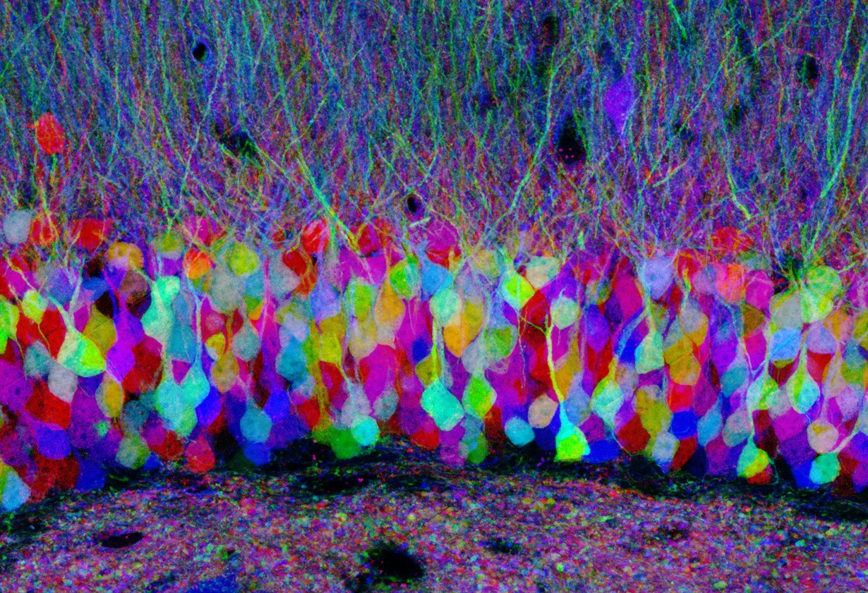
This specialized microscope allows us to view specific cellular structures through the use of fluorescently labeled molecules. Through genetic manipulation or chemical reactions, we are able to attach a fluorescent molecule (protein or organic molecule) to specific proteins. This fluorescent molecule is able to fluoresce a certain color if it is hit by light of a proper wavelength. A fluorescence microscope contains a light source that emits a light of a certain wavelength/energy that hits the sample that has the fluorescent molecules. The sample is then able to emit a light of a particular wavelength, and that light can be captured on a camera. This is a powerful tool to investigate where proteins are located in the cell, and gives us some insight to their role in the cell.

Confocal image of pastel neurons in the hippocampus of a "Brainbow" mouse brain, with each neuron expressing a distinct color. In Brainbow mice, neurons randomly choose combinations of red, yellow and cyan fluorescent proteins, so that they each glow a particular color. This provides a way to distinguish neighboring neurons and visualize brain circuits. Honorable Mention, 2007 Olympus BioScapes Digital Imaging Competition. For additional details see: Livet J, Weissman TA, Kang H, Draft RW, Lu J, Bennis RA, Sanes JR, Lichtman JW. Nature. 2007 Nov 1;450(7166):56-62. (CIL:42753)
